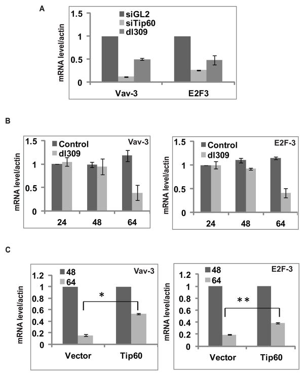
Figure 7. Tip60 degradation by adenovirus decreases Tip60 dependent cellular gene expression.
(A) Relative expression of cellular genes in MCF10A cells transfected with siGL2, siTip60 or infected with dl309. Cells were harvested 64 hrs post infection or transfection. Relative expression of genes by RT-qPCR compared to control siGL2. Mean ± SD (n=3).
(B) Decrease in cellular gene expression coincides with Tip60 degradation in dl309- infected cells. MCF10A cells were infected with mock or dl309 virus and cells were harvested at different time points as indicated. Relative mRNA expression of Vav-3 and E2F-3 was measured at indicated times by RT-qPCR taking value of Control 24 hrs sample as 1. Mean ± SD (n=3).
(C) Tip60 overexpression can rescue decrease in cellular genes expression. Relative mRNA expression of Vav-3 and E2F-3 at 48 and 64 hrs post infection in cells stably transfected with empty vector or Tip60 expressing plasmid. Level of expression at 48 hrs in vector or Tip60 overexpressing cells is taken as 1. Mean ± SD, n=3. (*P<0.05and **P<0.05 by Student’s t test).

