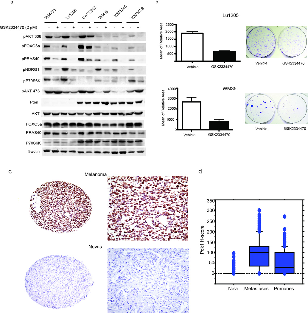
Figure 5. PDK1 inhibition attenuates AGC kinases in both Pten WT and mutant melanomas.
(a) Western blot analysis was performed using protein lysates from human melanoma cells with Pten mutation (793, Lu1205, UACC903) or Pten WT (WM35, WM1346, and WM3629) with the indicated antibodies. (b) Human melanoma cell lines Lu1205 and WM35 were treated with either vehicle or GSK2334470 (2.5 µM, changing the media every other day) and monitored for colony formation assay. The graph represents the quantification of the mean relative area after 7 days (Lu1205) and 12 days (WM35) in culture (representative images of colonies grown in culture are shown on the right panel). Analysis was performed in triplicates and repeated two times. Error bars represent SEM. (c) Expression of PDK1 was studied in a large cohort of primary and metastatic melanomas and in nevi. Examples of strong and weak staining are shown. (d) Box plots demonstrate differences in expression in the three categories of samples, with the PDK1 H-score (scale 0–300) on the Y-axis. Expression was significantly lower in nevi and primary lesions than metastases (P = 0.0001).

