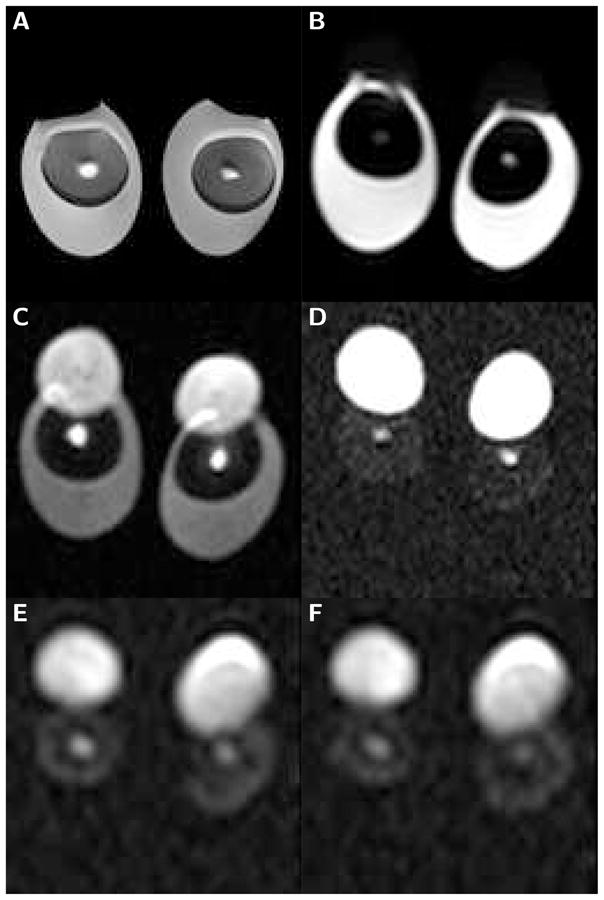
Figure 1.

Sub-images (100 mm field of view) of boiled and raw egg-pairs scanned with the localizing sequence and LSDI. The brightness of the individual images has been adjusted for optimal viewing. A) Spin-echo image (TE=30 ms) of eggs boiled for 5 minutes. B) High-resolution line scan diffusion image (b=5 s/mm2) of raw eggs. C) High-resolution line scan diffusion image (b=2,000 s/mm2) of raw eggs. D) High-resolution line scan diffusion image (b=5,000 s/mm2) of raw eggs. E) Low-resolution line scan diffusion image (b=20,000 s/mm2) of raw eggs. F) Low-resolution line scan diffusion image (b=50,000 s/mm2) of raw eggs. The latebra is clearly visible as a bright structure in the center of the yolk. It evidently maintains signal above noise, even at a b-value of 50,000 s/mm2. In contrast, already at 5,000 s/mm2 the water signal of the egg white falls well below the noise threshold. On the diffusion-weighted images the lipid containing portion of the yolk is shifted upwards, permitting an unencumbered water diffusion analysis of the latebra.
