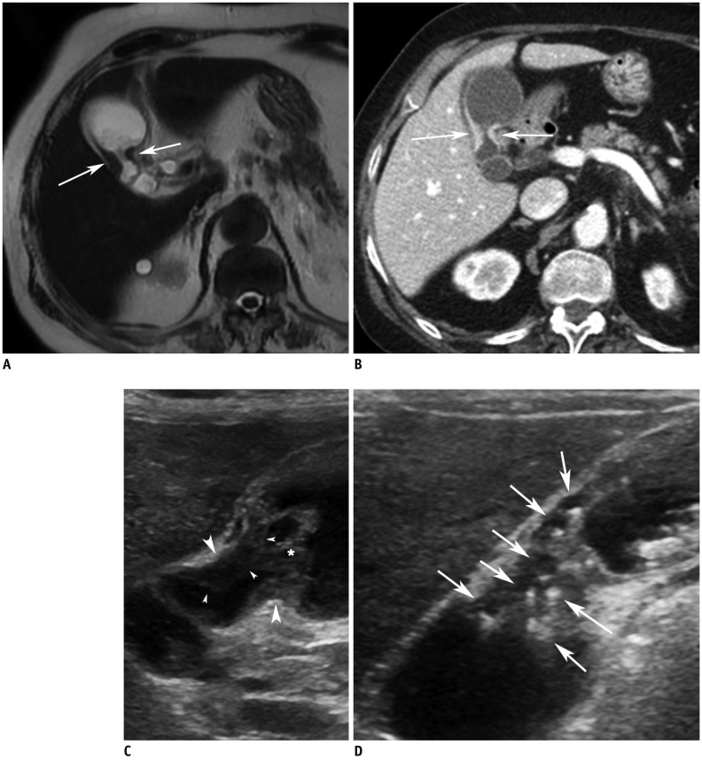
Fig. 2.
Gallbladder body cancer (pT2) in 81-year-old female.
A. MR image with T2-weighted single-shot fast spin-echo sequence shows segmental thickening (arrows) without evidence of intramural cysts at body of gallbladder. B. CT image during portal venous phase shows segmental wall thickening with strong contrast enhancement at body (arrows). C. High-resolution ultrasound image showing segmental wall thickening (arrowheads) without evidence of intramural cysts or intramural echogenic material deposition at body. Asterisks (*) indicate sludge. D. High-resolution ultrasound image in patient with segmental adenomyomatosis shows multiple cysts and echogenic materials (arrows) within thickened walls. This image clearly shows difference between adenomyomatosis and cancer on ultrasound image.

