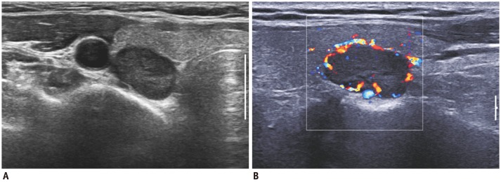
Fig. 18.

Parathyroid adenoma in 54-year-old woman.
A. Thyroid US revealed hypoechoic solid mass at right lower pole region. B. Color Doppler US showed characteristic peripheral vascularity. It was surgically shown to be parathyroid adenoma. US = ultrasonography
