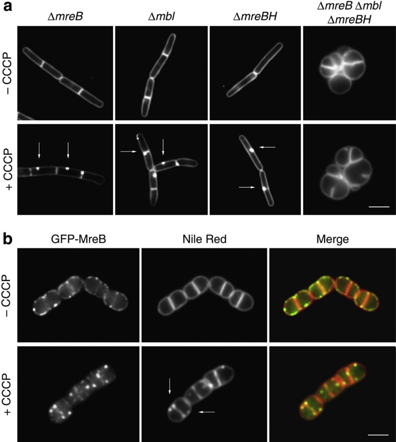
Figure 2. Distorted lipid staining requires the MreB cytoskeleton.
(a) CCCP-dependent fluorescent Nile Red foci occur in B. subtilis strains deficient for single MreB homologues, but are absent in strains that lack all three MreB homologues (ΔmreB, Δmbl, ΔmreBH). Some of the Nile Red foci appearing with CCCP are highlighted with arrows. See also Supplementary Fig. 7 for the analysis of the foci frequency. Strains used: B. subtilis 3728 (ΔmreB), 4261 (Δmbl), 4262 (ΔmreBH) and 4277 (ΔmreB, Δmbl, ΔmreBH). (b) Colocalization of GFP-MreB (left panels) with Nile Red (middle panels) in B. subtilis cells depleted for RodA. The MreB cytoskeleton is disrupted with CCCP. Some of the Nile Red foci appearing with CCCP are highlighted with arrows. Strain used: B. subtilis HS36 (gfp-mreB Pspac-rodA). Scale bar, 2 μm.

