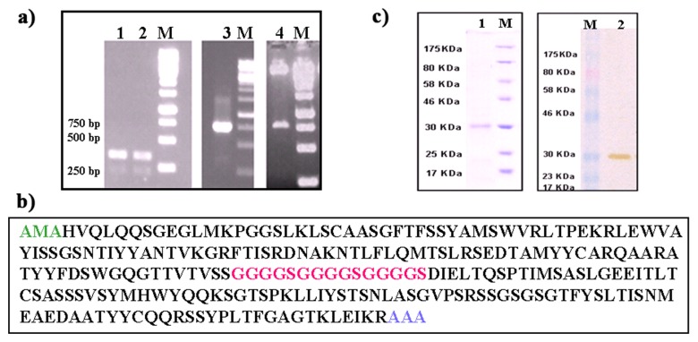
Fig. 1.
(a) Electrophoretic analysis of PCR amplified variable heavy and light chain domains. These gene sequences were amplified from total RNA isolated from a hybridoma secreting cell line IL-A71. Lane M: Molecular weight markers. Lanes 1, 2, 3 show variable heavy and light chain genes and assembled scFv PCR products. Lane 4 shows the release of 750 bp product, recombinant expression cassette after EcoRI and NotI digestion. (b) Amino acid sequence of anti-bovine IgA scFv containing VL, linker peptide and VH. The linker peptide is marked in italics. Underlining indicates the restriction enzyme sites for cloning the scFv gene. (c) SDS-PAGE and Western blot analysis of scFv expression. Lane M: molecular mass marker. Lanes 1 and 2 are purified samples from Ni-NTA agarose column. Sample was separated with 12% SDS-PAGE, followed by Coomassie blue staining (lane 1). For Western blotting of purified sample from Ni-NTA affinity column was transferred onto a PVDF membrane and probed with anti-His probe for 1 h. The antibody was detected by using DAB substrate (lane 2)

