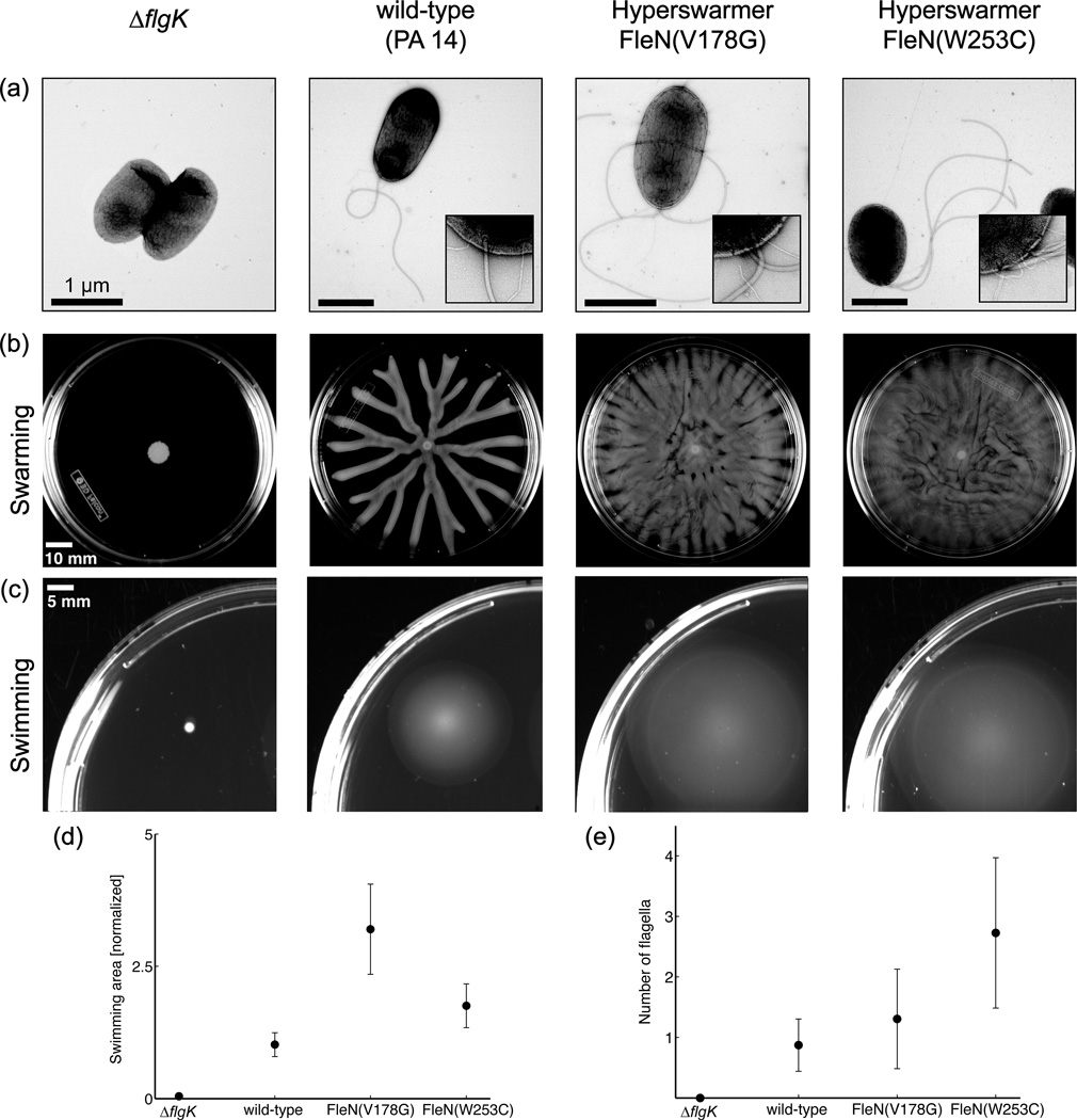
Figure 1.

Motility patterns of non-flagellated (∆flgK), mono-flagellated (wild-type) and multi-flagellated (two hyperswarmer clones) Pseudomonas aeruginosa, as shown by transmission electron microscopy (a). Insets show 88,000× magnification of the cell pole. Swarming (b) and swimming (c) motility are dependent on the number of flagella. (d) Quantification of the swimming area for the different strains and (e) quantification of the number of flagella. Data and pictures reproduced from van Ditmarsch14.
