Figure 5.
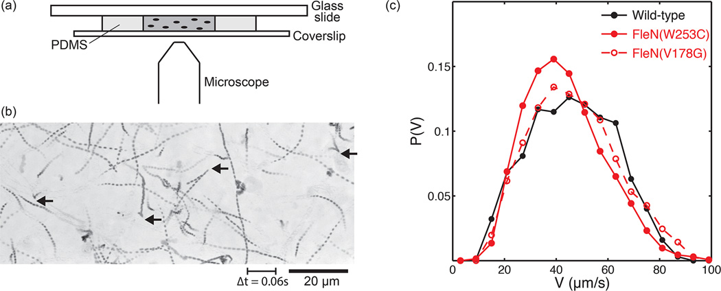
Swimming of P. aeruginosa in phase contrast microscopy: (a) Diagram of PDMS microchamber used for imaging. (b) Minimum intensity temporal projection image reveals individual trajectories, made of nearly straight runs and sharp turns (indicated with black arrows). (c) Speed distributions show that wild-type swims slightly faster than the two hyperswarmers.

