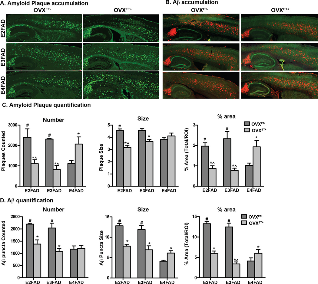
Figure 1. Extracellular amyloid plaque and Aβ deposition is decreased with APOE2 and APOE3 but increased with APOE4, in ovariectomized EFAD mice treated with estradiol.
Representative image of sagittal sections from ovariectomized EFAD mice treated with vehicle (OVXET−) or estradiol (OVXET+), and A. stained with Thio-S or B. immunostained for Aβ (red) and NeuN (green) (x10 magnification). Quantification of number, size and % area covered in the frontal cortex of C. Thio-S stained plaque plaques or D. extracellular Aβ. Data are expressed as the mean ± S.E.M, and were analyzed by one-way ANOVA followed by Tukey’s multiple comparison post hoc analysis. n = 5–6. *p < 0.05 OVXET− versus OVXET+. #p < 0.05 vs E4FAD for OVXET−, ^p < 0.05 vs E4FAD for OVXET+

