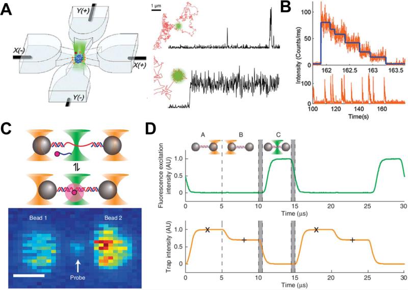
Fig. 2.
(A) Schematic of the ABEL trap, where a single molecule is trapped in the center of a four-channel microfluidic chamber. Each channel is connected to an electrode to provide bias electric field generating electric kinetic forces to balance the Brownian motion. A well trapped single fluorescent molecule can provide consistent fluorescent signals to be collected using a confocal fluorescent microscope. (B). Lower spike fluorescent signals are from Cy3-APD interactions. The generations of those signals indicate the interactions between a trapped TRiC using ABEL and the nucleotides entering the trap. Each spike fluorescent signal is then photobleached with stepwise bleaching event shown in the upper part of the figure. (C) Schematic setup and fluorescent images of dual optical traps with ultra-high precision and resolution. The scale bar is 1 μm. (D) To avoid excessive heat generated during trapping to harm the single molecules, trapping lasers and excitation light are sequentially turned “ON” and “OFF” in a programmable manner that is controlled digitally. This trapping method also offers high-resolution detection and better fluorescence lifetime. Images are reproduced from Ref. 51, 52, and 55.

