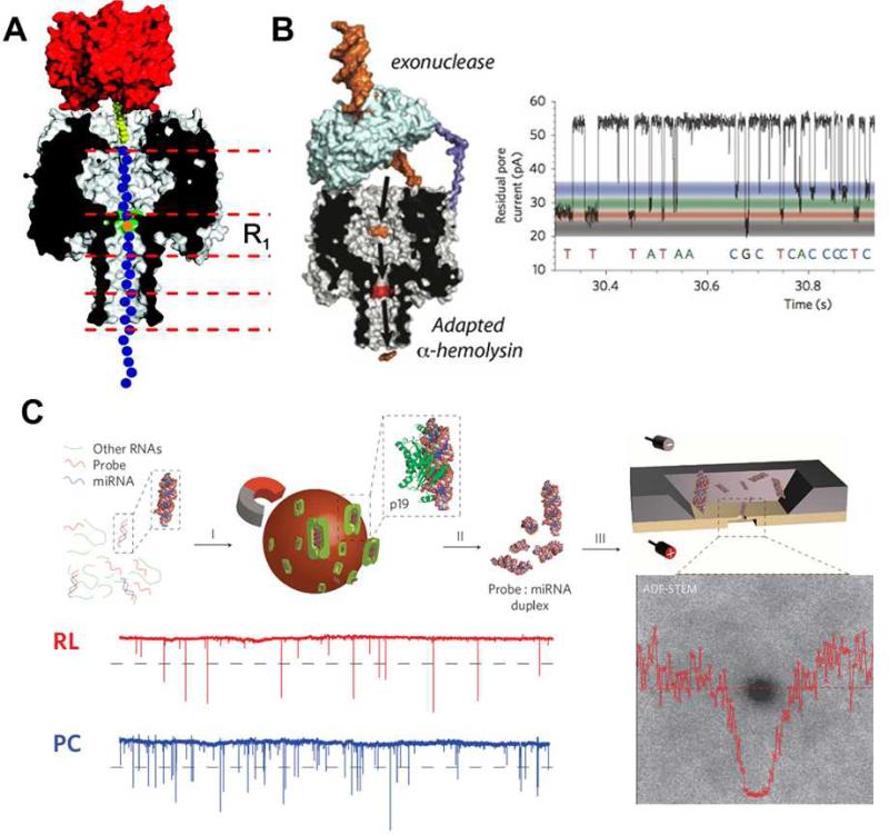
Fig 6.
(A) Immobilization of a ssDNA (blue) in an α-hemolysin nanopore (grey/black) by the interaction between the biotinylated (yellow) end of the DNA strand and streptavidin (red). The intensity of nucleotide (orange) within the strand that resides at the R1 position (green) can be identified by its characteristic electric fingerprint. (B) Scheme of exo-sequencing and residual pore current recording from the α-hemolysin nanopore attached to a cyclodextrin adapter. The 4 nucleoside monophosphates in the solution can be discriminated by the characteristic residual current levels. (C) Scheme of the ultra-thin nanopore based miRNA sensing and quantification method. The miRNA enriched sample (RL) gave comparable spike-like pulse signals to those of the positive control (PC). Images are reproduced from Ref. 22, 85, and 89.

