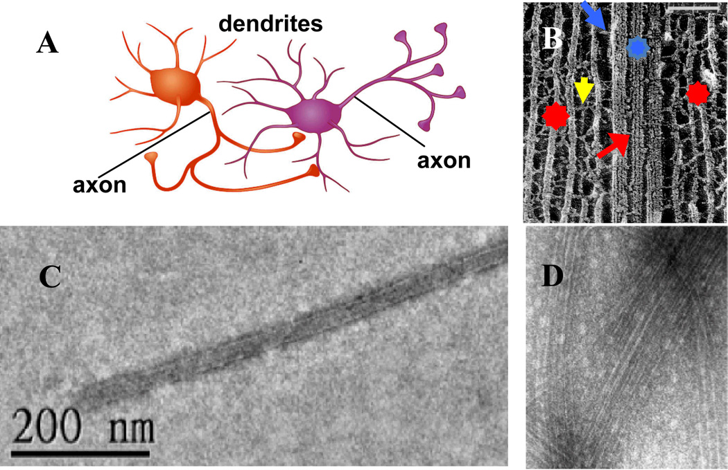
Fig. 1.
(A) Two interacting neurons with orange neuron synapsing on the body and dendrite of purple neuron. (B) Cryo-Electron micrograph (deep-etched) of an axonal cytoskeleton in a spinal cord vertebrate neuron. In axons, neurofilaments (NFs, red stars), are the majority component and form oriented extended arrays surrounding microtubule (MT) bundles (blue star) or single MTs. Cross-bridges between NFs (NF-sidearm, yellow arrow), MTs (MAP tau, red arrow) and MT and NF-sidearms (blue arrow) are evident. In B Bar is 100 nm. (C) TEM of taxol-stabilized MT (taxol:tubulin, 1:1) coated with MAP tau (3RS isoform) showing lack of bundle formation even for a high tau/tubulin molar ratio of 0.5 (D) TEM of taxol-stabilized MT (taxol:tubulin, 1:1) coated with MAP tau (4RS isoform) showing evidence of loose bundles at a very high tau/tubulin molar ratio of 1.7. Bar in D is 100 nm. (B) Adapted from reference 6. © N. Hirokawa,1991.

