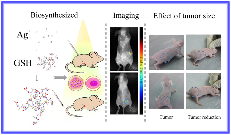
Figure 5. Representative xenograft tumor nude mice models of Cervical carcinoma observed in normal light (indicating tumor size).
The [Ag(GSH)]+ complex solution was administered into the solid tumor mouse model through local injection or intravenous injection through the tail. After incubation for 24 h, fluorescent silver nanoclusters were observed inside the tumors by in vivo fluorescence imaging using a 590 nm excitation wavelength. After 7 days the corresponding tumor was reduced in size and eventually disappeared. In comparison, the negative control groups, which received an equivalent volume of Phosphate Buffered Saline (PBS), did not exhibit any apparent fluorescence. (This figure was prepared by using open source software of Photoshop. The left panel is a cartoon illustration drawn by co-authors for the relevant experimental procedure, including the complexes of Ag+ with GSH and then biosynthesized to Ag NCs for in vivo bio-imaging study, while the middle and right panel is the experimental observations for in vivo tumor bio-images, where the [Ag(GSH)]+ complex solution was administered into the solid tumor mouse model through local injection or intravenous injection through the tail.).

