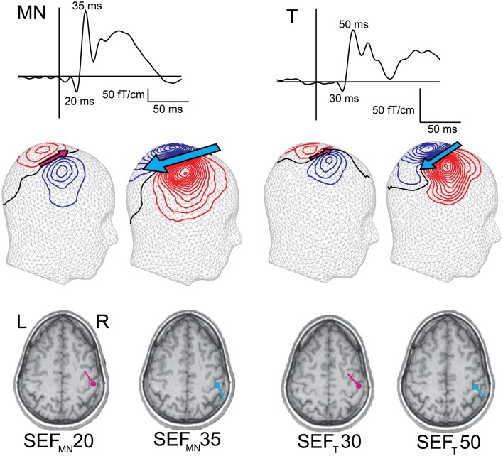
Figure 2.
Early adult SEFs to median nerve (SEFMN) and tactile (SEFT) stimulation. The SEF waveform from one gradiometer channel shows SEFMN20/SEFT30 and SEFMN35/SEFT50 deflections, and the magnetic contours – reflected on a head surface – reveal the different current orientations (indicated by the pink and blue arrows). Compared with SEFMN, the SEFT latencies are slightly longer and amplitudes lower (note the different amplitude scales). Both stimulation methods, however, elicit an initial anteriorly pointing dipolar source, SEFMN20/SEFT30 (though in some subjects this deflection is minute to tactile stimulation), followed by a posteriorly pointing dipolar source, SEFMN35/SEFT50. The dipoles are superimposed on individual MRIs and both sources are localized in SI.

