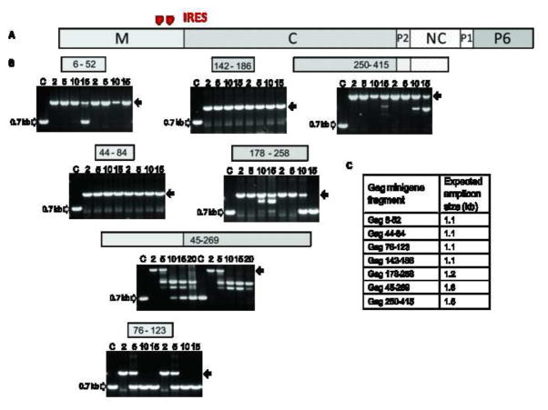
Figure 2.
Viral genetic stability studies performed in seven recombinant YF 17D viruses expressing different Gag fragments. (A) Organization of SIV gag gene coding Matrix (M), Capsid (C), Nucleocapsid (NC) proteins and the carboxy-terminal domain P6 as well as the spacer regions P1 and P2 (GenBank AY588946). (B) Electrophoretic analysis of amplicons containing the insertion region obtained by RT-PCR of viral RNA extracted from supernatant of cultures after serial passages in Vero cells. Numbers of passages are indicated above the figures. Grey arrows indicate the size of amplicons for viruses without insertion. Black arrows indicate the expected amplicon size for each recombinant virus containing the complete insertion. The corresponding Gag fragment that is expressed by each recombinant YF virus is specified above the electrophoretic profile. (C) Expected amplicon sizes for each recombinant virus.

