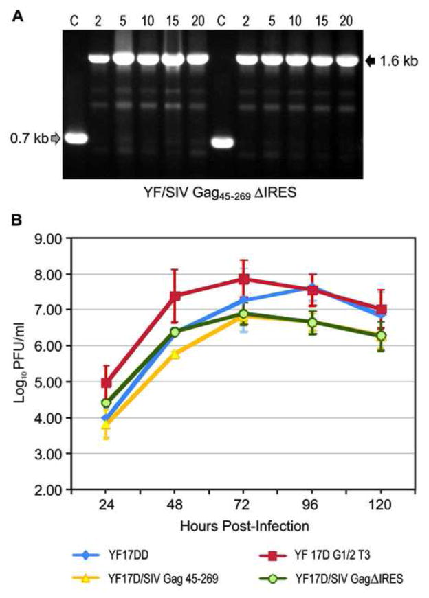
Figure 4.
Viral genetic stability and proliferation studies of the new recombinant YF17D/SIV GagΔIRES virus. (A) Electrophoretic analysis of amplicons containing the insertion region obtained by RT-PCR of viral RNA extracted from supernatant of cultures after serial passages in Vero cells. Numbers of passages are indicated above the figure. On the right, a black arrow indicates the size of the amplicon containing the complete heterologous expression cassette. On the left, the gray arrow points to the position of the YF 17D vaccine amplicon that does not contain any insertion. (B) Viral growth curves in Vero cells. Cells were infected with either the control YF 17DD (vaccine virus strain) and YF17D/G1/2T3 (parental clone) viruses or the recombinant YF17D/SIV Gag45-269 or YF17D/SIV GagΔIRES viruses at MOI of 0.02. Each time point represents the average titer obtained from three independent experiments with the respective standard deviations.

