
Figure 3.
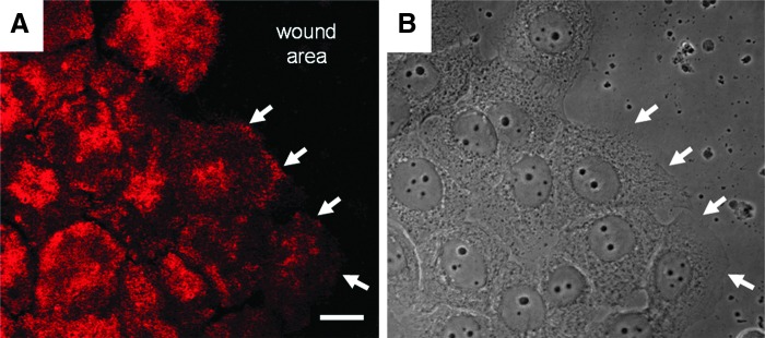
Immortalized human keratinocytes were allowed to attain confluence in vitro and were then scratched wounded. At 6 h after wounding, the cells were prepared for confocal immunofluorescence using antibodies against β4 integrin (A). Cells at the wound edge have begun to move onto the wound site. White arrows indicate β4 integrin staining along the leading lamellipodia of cells at the wound margin. These same areas are marked by arrows in the phase image of the cells in (B). Scale bar, 20 μm.