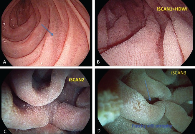
Figure 1).

High-definition iScan (Pentax, Japan) technique showed patchy irregularity of the mucosa in the second portion of the duodenum. A Normal white-light endoscopy showing normal second part of duodenum. B to D iScan virtual chromoendoscopy high-definition in combination with water immersion technique with virtual chromoendoscopy showing transitional patchy area of normal and atrophic villi. Biopsies confirmed celiac disease
