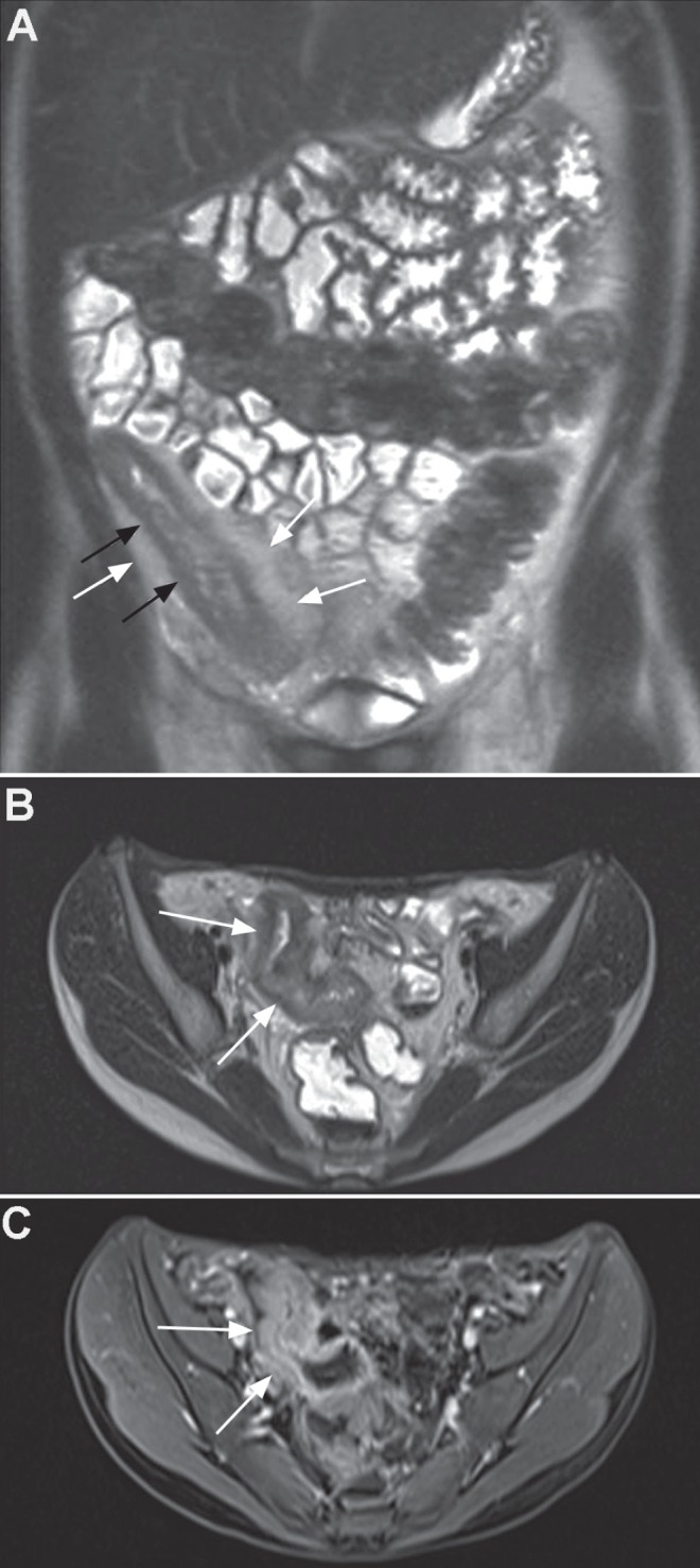
Figure 1).

A Coronal T2 half-Fourier acquisition single-shot turbo spin-echo (HASTE) image showing colonic and particular terminal ileal disease with bowel wall thickening (black arrows) and fibrofatty proliferation (white arrows). B Axial T2 HASTE image showing striking distal ileal bowel wall thickening (arrows). C Contrast-enhanced volumetric interpolated breath-hold sequence showing enhancement of the thickened bowel wall (arrows)
