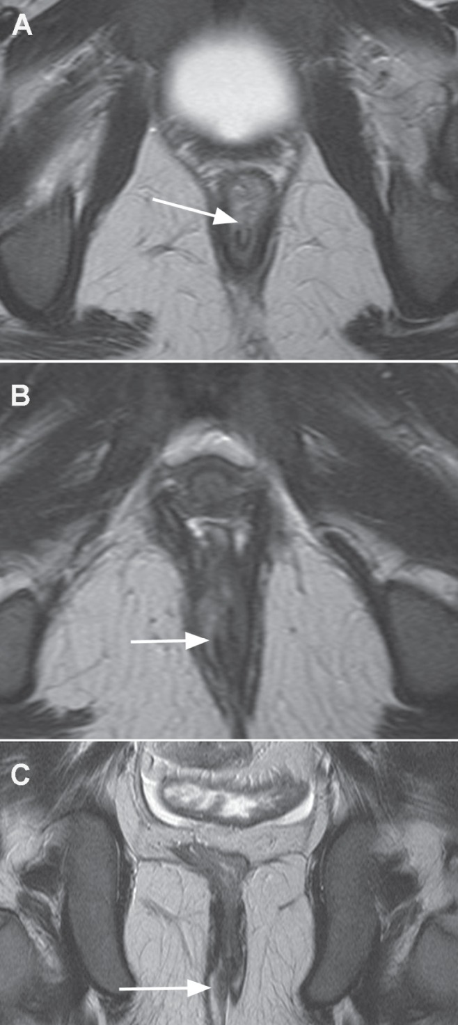
Figure 5).

A Upper axial T2 image demonstrating the start of the fistula breaching the internal sphincter at 12:00 (arrow). B Mid axial T2 image demonstrating the fistula between internal and external sphincters (arrow). C Coronal T2 image demonstrates inferior extent of the trans-sphincteric fistula breaching through the external sphincteric muscles (arrow)
