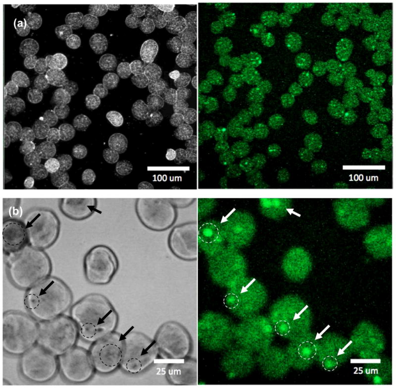
Figure 7.

Confocal microscopy (left) gray and (right) fluorescent images of MEAN loaded magnetic alginate microspheres (λexcitation=445 and λemission=550 nm) ((a) low magnification and (b) high magnification, arrows indicate punctate regions of condensed USPIO clusters and intense fluorescent signal).
