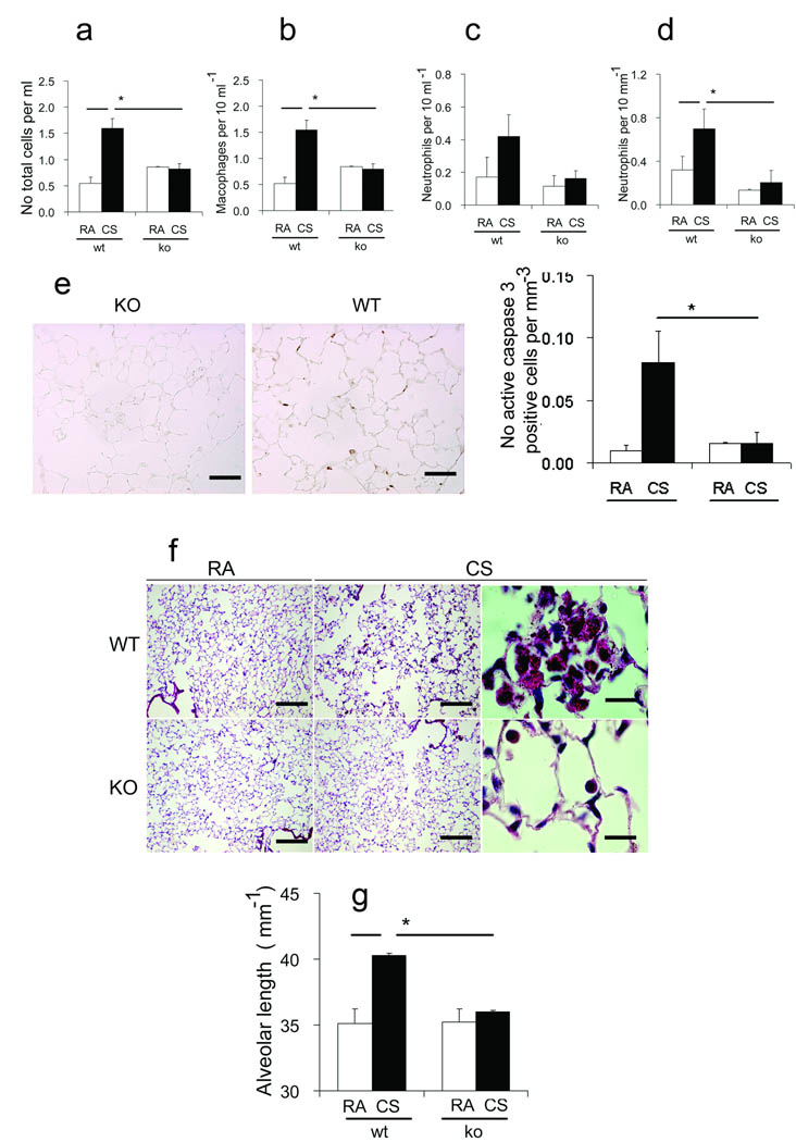
Figure 5. Rtp801 − / − mice are protected against cigarette smoke – induced pulmonary inflammation, apoptosis, and emphysema.
Bal total cell counts (a), including numbers of macrophages (b), and neutrophils (c), infiltrating neutrophils in alveolar lung tissue (d) (µm−1 alveolar length), active caspase 3 – positive cells (brown, arrows) (upper) (× 50 µm) and numbers of active caspase 3 – positive cells (µm−1 alveolar septa) (lower) in Rtp801 wildtype and Rtp801 − / − mice kept in RA or exposed to CSk for 7 days (n = 3 and 7 mice, respectively). Alveolar morphology in Rtp801 wildtype CSk for 6 months (f) showing airspace enlargement and large clusters of alveolar macrophages containing smoking pigment in the cytoplasm when compared with Rtp801 − / − mouse lungs (× 250 µm, right and middle; × 25 µm, left). Alveolar length represent mean linear intercepts (g) (n = 5 to 7 mice in each group). *: P < 0.05

