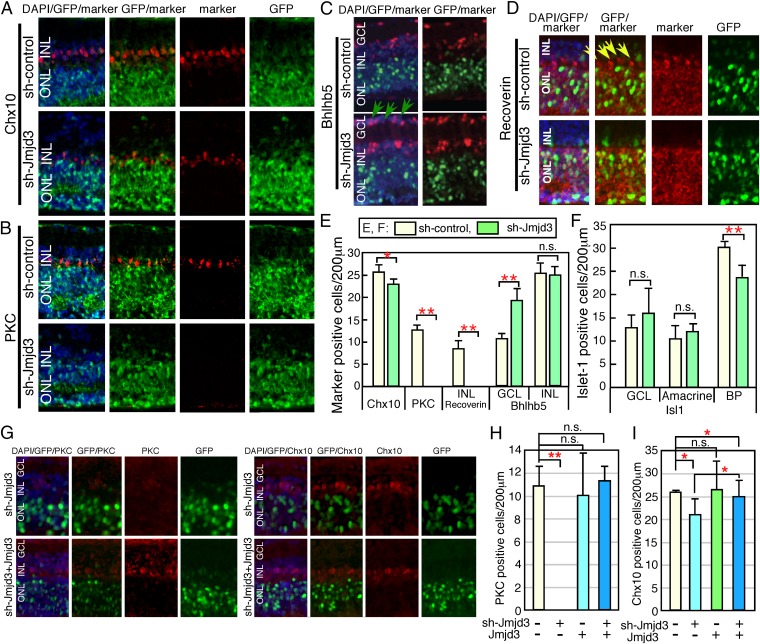
Fig. 2.
Knockdown of Jmjd3 expression during retinal development. Plasmids encoding CAG-EGFP/U6-shRNA-Jmjd3 (sh-Jmjd3)/control U6 (sh-control) (A–D), or CAG-EGFP/sh-Jmjd3/Jmjd3 expression plasmid (G–I) were introduced into the retina (E17) by electroporation. After 2 wk of explant culture, the retina was frozen-sectioned and immunostained using antibodies to antiretinal subset markers as indicated. In E, F, H, and I, the number of marker-positive cells in the electroporated region (200 μm) as judged by EGFP fluorescence was counted in sections. Green arrows in C indicate displaced amacrine cells, and yellow arrows in D indicate cone-OFF-BP cells. More than five sections from three independent samples were counted, and values with SDs are shown. **P < 0.01, *P < 0.05, and P > 0.05 (n.s.) were calculated by the Student t test. n.s., not significant.

