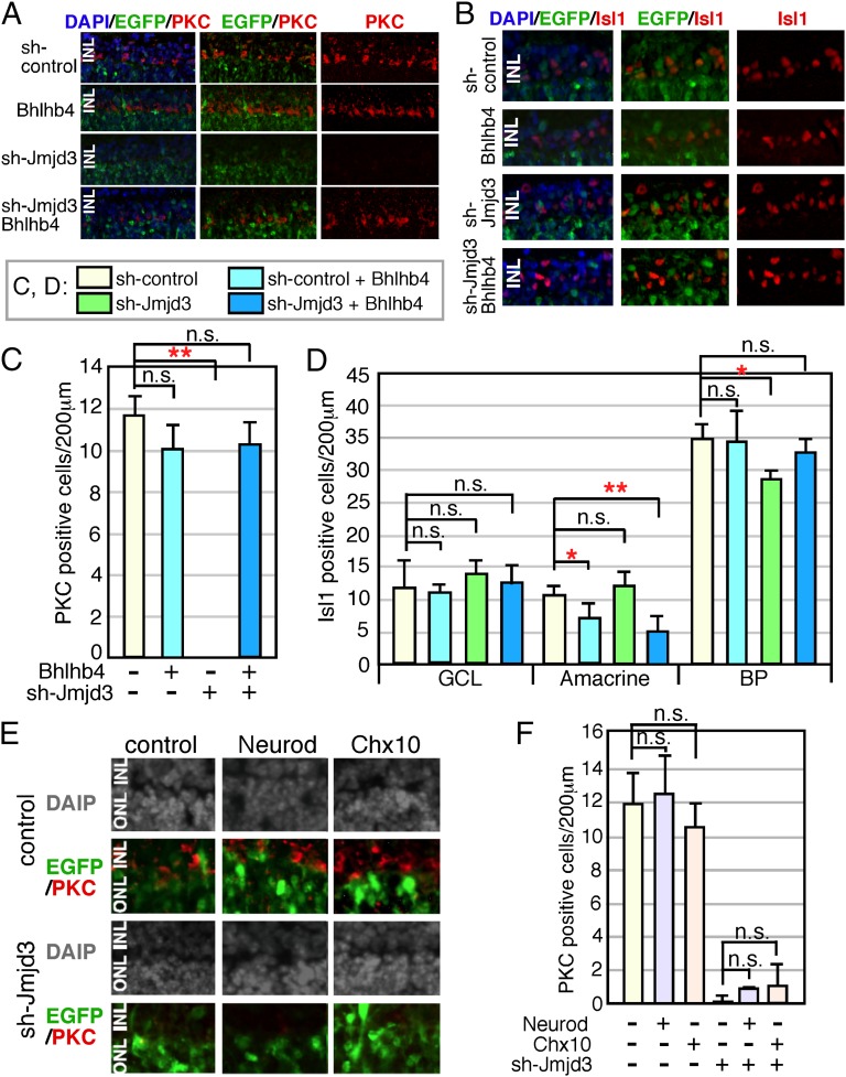
Fig. 4.
Supplemental expression of Bhlhb4 into sh-Jmjd3–transfected retina. Mouse retina at E17 was isolated, and plasmids encoding CAG-EGFP and sh-Jmjd3 or sh-control with or without CAG-Bhlhb4 (A–D) and CAG-Neurod or CAG-Chx10 (E and F) were introduced into the retina by electroporation. The total amount of transfected plasmids was adjusted as the same for all experiments by the addition of empty vector. (A, B, and E) After 2 wk of explant culture, the retina was frozen-sectioned and immunostained using antibodies to antiretinal subset markers as indicated. (C, D, and F) Number of marker-positive cells in the electroporated region (200 μm) was counted. More than five sections from three independent samples were counted, and values with SDs are shown. **P < 0.01, *P < 0.05, and P > 0.05 (n.s.) were calculated by the Student t test.

