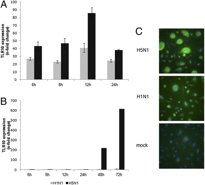
Fig. 2.
Kinetics of influenza A virus-induced TLR10 expression in human macrophages. Expression of TLR10 in human macrophages infected by H1N1 or H5N1 influenza A viruses at (A) MOI of 2 or (B) 0.001 compared with mock infection at different postinfection time were assessed by RT-PCR. The result of one representative experiment from three independent experiments with three donors is shown. Error bars indicate SD of three technical triplicates. (C) TLR10 protein expression (FITC-green) was detected using immunofluorescent staining. Cells were counterstained with DAPI (blue) and viewed in a fluorescent microscope (magnification 400×). Multinucleate giant cells are seen, especially in H5N1 virus infected cells.

