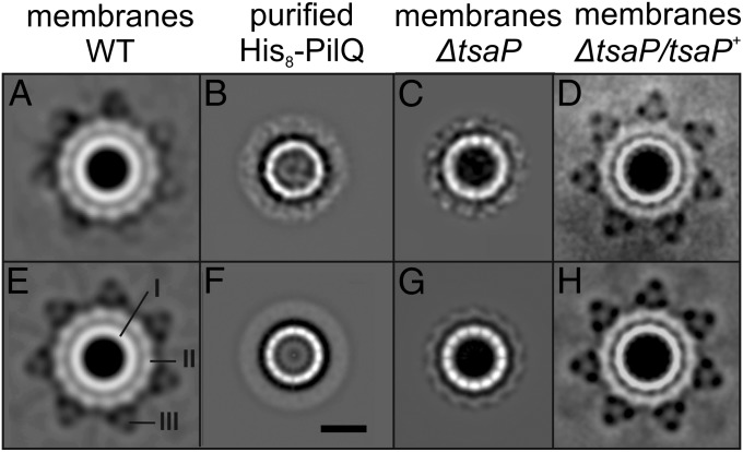
Fig. 1.
Projection maps of single-particle EM analysis of the PilQ complex from N. gonorrhoeae. Projection maps of class averages of single-particle EM images obtained from membranes isolated from the WT (A and E), the ΔtsaP strain (C and G), and the ΔtsaP/tsaP+ strain (D and H) grown in the presence of 1 mM IPTG are shown. (B and F) Class averages of single-particle EM images of the solubilized and purified His8–PilQ complex. Projection maps without (A–D) and with (E–H) 14-fold imposed symmetry are depicted. I, II, and III indicate the inner ring, the peripheral ring, and the spikes, respectively. (Scale bar: 10 nm.)

