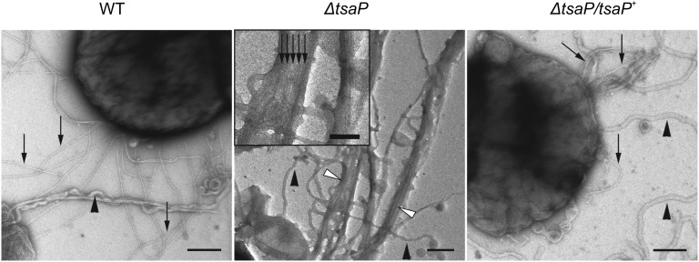
Fig. 4.
Deletion of TsaP leads to formation of membrane protrusions containing T4P in N. gonorrhoeae. An EM analysis of WT, ΔtsaP, and ΔtsaP/tsaP+ strains grown in the presence of 1 mM IPTG was performed. Cells were applied to carbon-coated copper grids, washed twice with double-distilled water, and subsequently stained with uranyl acetate before investigation via EM. T4P (black arrows) and membrane blebs (black arrowheads) are shown. (Inset) Membrane protrusions (white arrows) observed in the ΔtsaP mutant are filled with T4P. (Scale bars: main images, 200 nm; Inset, 50 nm.)

