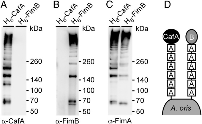
Fig. 4.
Distinct fimbrial polymers formed by CafA and FimA independent of FimB and FimA polymers. Cell wall extracts of A. oris strain ΔcafA or ΔfimB expressing CafA or FimB, respectively, with a “6×-His tag inserted upstream of the LPXTG motif, were used for affinity chromatography. Purified proteins were subjected to immunoblotting with α-CafA (A), α-FimB (B), or α-FimA (C). (D) Schematic representation shows that A. oris assembles two distinct fimbrial structures made of FimA, forming the fimbrial shaft, and CafA or FimB, each constituting the fimbrial tip.

