
Figure 7.
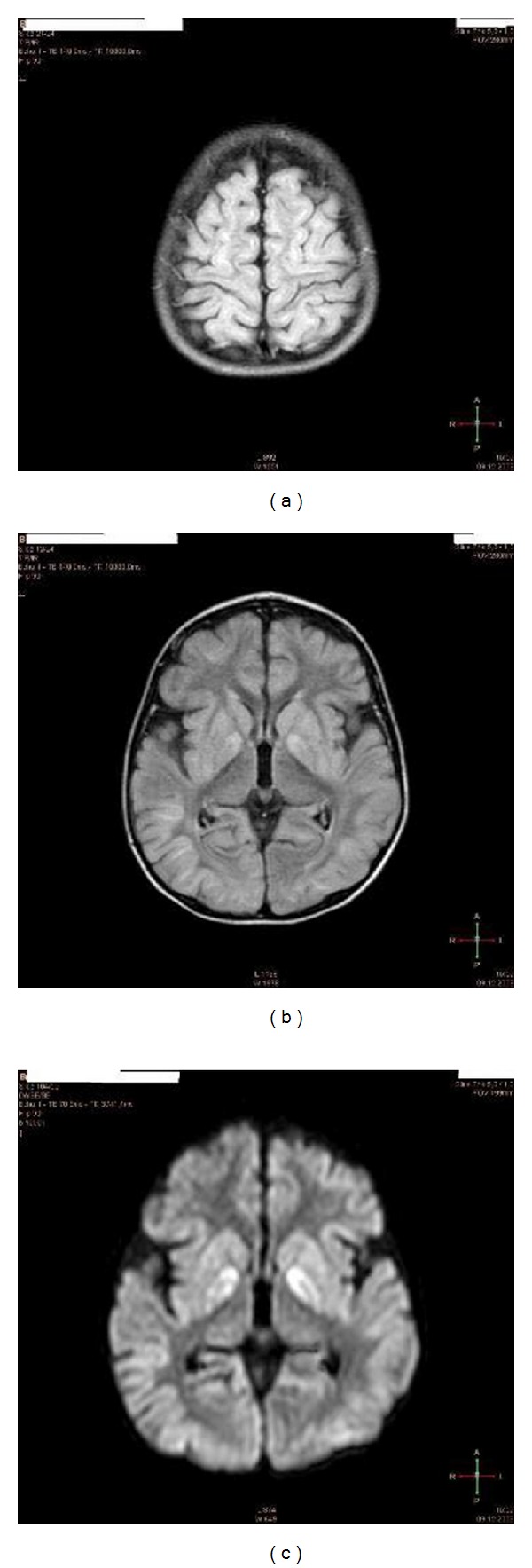
Brain MRI of the patient (L. B.) 2 weeks after cardiac arrest. Note, signs of severe global ischemia in cortical structures as evidenced by (a) signal hyperintensity of gyri in almost entire cortex (FLAIR sequences) and basal ganglia (b), including caudate nucleus and putamen (c) (FLAIR DWI sequences with contrast media) (see video S1, S2 in Supplementary Material [44]).