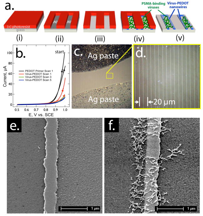
Figure 2.
Synthesis of virus-PEDOT nanowires: a) Process flow for the preparation, using the LPEN process, of virus-PEDOT composite nanowires for the detection of PSMA. b) Cyclic voltammograms for the electrodeposition of virus-PEDOT nanowires within the LPNE microfabricated template. Two primer scans were first carried out in aqueous 2.5 mM EDOT, 12.5 mM LiClO4 (black). Then five additional scans in 2.5 mM EDOT, 10 nM PSMA-binding viruses, and 12.5 mM LiClO4 were used to build up a virus-PEDOT composite nanowire 200–300 nm in total width. c, d) Optical micrographs of the resulting nanowire array on glass after the application of silver paste electrical contacts, e, f) Scanning electron micrographs of single nanowires of pure PEDOT (e), and the virus-PEDOT composite (f). The net-like structures observed in (f) are aggregates of filamentous M13 virus particles.

