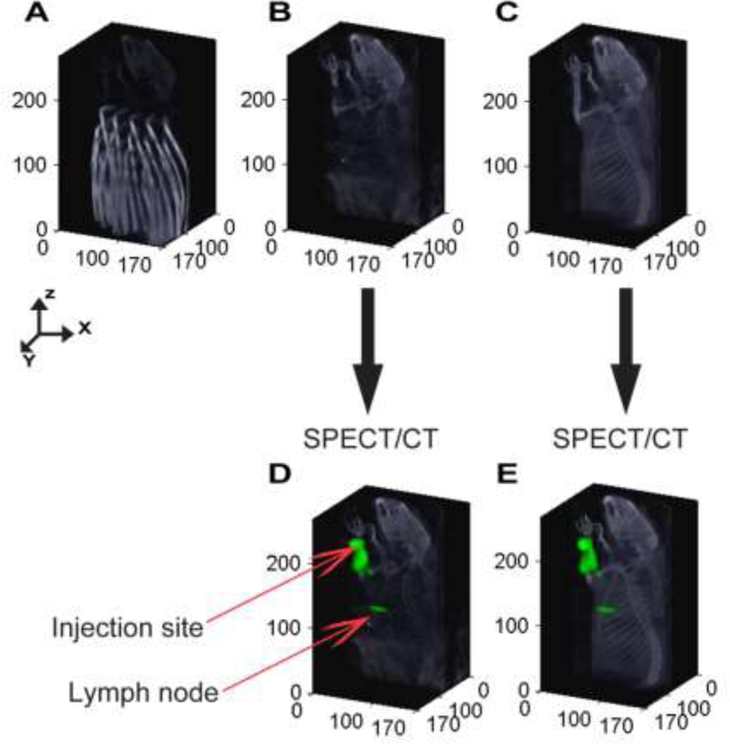
Figure 2.
Evaluating the influence of fiber arrays on SPECT and CT images: (A) X-ray CT image of a rat with the fiber array of DOT. (B) X-ray CT image of the same rat shown after removing the voxels related to the fiber array of the DOT. (C) X-ray CT acquired after removing the DOT imaging pad. The anatomical structures acquired without the fibers are used to display the fluorescence and radioactive distribution for all 5 rats. (D) SPECT data acquired at the presence of DOT fiber array depth in a rat following injection of multimodal imaging agent, 111In-BS255, into the left forearm. (E) SPECT data of the same rat acquired after removing the fibers.

