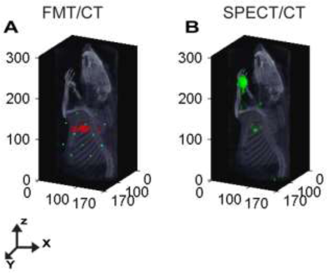
Figure 3.
Representative Sentinel Lymph Node Mapping using Optical and Nuclear Imaging Systems. (A) DOT-CT image of the fluorescent LNs shown at 2mm depth in a rat following injection of multimodal imaging agent, 111In-BS255, into the left forearm. (B) SPECT-CT image demonstrating localization of axillary lymph node identified by accumulation of the multimodal imaging agent.

