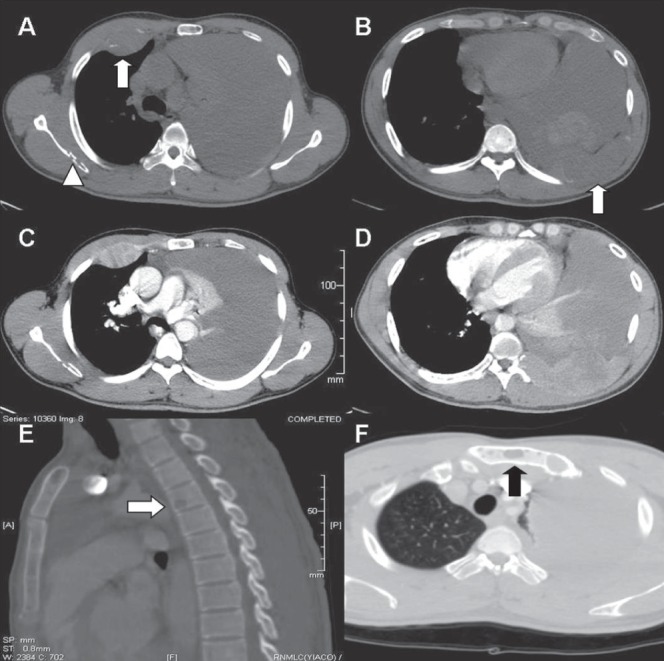
Figure 2).

A Plain axial computed tomography (CT) image in the bone window demonstrates a soft tissue mass in the anterior aspect of the right fourth rib (arrow) and an area of osteolysis within the right scapula (arrow head). Massive unilateral pleural effusion is also noted. B Axial plain CT image at a lower level shows a destroyed left ninth rib posteriorly with soft tissue component (arrow) and areas of hyperdensity within the effusion in keeping with hemorrhagic nature. C and D Heterogenous postcontrast enhancement of the soft tissue masses is demonstrated. E A sagittally reformatted CT in the bone window demonstrates a lytic lesion in the body of the T4 vertebra (arrow). F Another lytic lesion is seen in the manubrium of the sternum (arrow)
