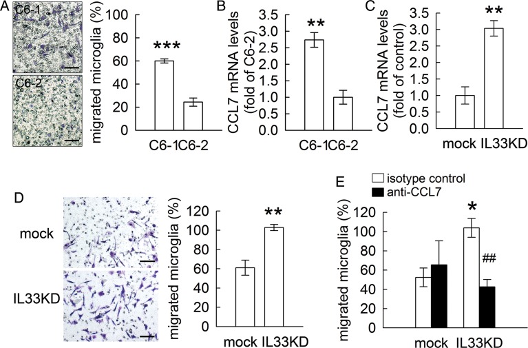
Fig. 11.
C6-derived CCL7 involvement in enhanced invasion of microglia. (A). Primary rat microglia were replated onto the upper side of the Transwell filter and then co-cultured with C6-1 or C6-2 cells. After 24 hours, the cells that migrated to the bottom side of the filter were stained (left-handed panel) and counted. C6-1 cells displayed a greater invasive ability than C6-2 cells. (B). Total RNA isolated from C6-1 cells or C6-2 cells was subjected to Q-PCR for the measurement of CCL7 mRNA expression. (C). Total RNA isolated from C6-mock and C6-IL33KD cells was subjected to Q-PCR for the measurement of CCL7 mRNA expression. (D). Primary rat microglia were seeded onto the upper side of the Transwell filter and then co-cultured with C6-mock or C6-IL33KD. The cells that migrated to the bottom side of the filter were stained (left-handed panel) and then counted. (E). Microglia were replated onto the upper side of the Transwell filter and then co-cultured with C6-mock or C6-IL33KD in the presence of isotype control or anti-CCL7/MCP3antibodies for an 8 hour incubation. Microglia that invaded to the bottom of the filter were counted.Scale bar in A and D, 100 μm. Data are means ± SEM of 3 independent experiments. *P < .05; **P < .01; ***P < .001 versus C6-2 (A and B) or C6-mock (C, D and E); ##P < .01 versus isotype control.

