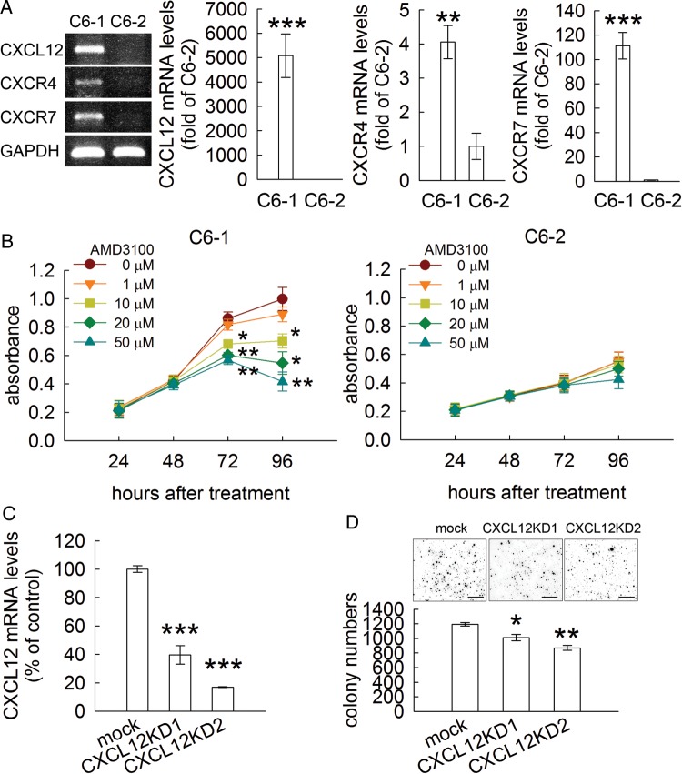
Fig. 3.
C6-1 cell growth suppressed by inhibition of CXCL12 action. (A). CXCL12, CXCR7, and CXCR4 mRNA levels in C6-1 and C6-2 cells were measured by Q-PCR. The 3 genes were expressed abundantly in C6-1 cells but not in C6-2 cells. Gel-based RT-PCR was used to verify the specificity of the primers for CXCL12, CXCR4, and CXCR7 (left-hand panel). (B) After treatment of C6-1 or C6-2 cells with AMD3100 (a CXCR4/CXCR7 inhibitor) for the various time periods indicated above, the cultures were subjected to MTT cell viability assay. AMD3100 at the concentrations greater than 10 μM effectively suppressed C6-1 cell growth but had no effect on the cell growth of C6-2 cells. (C) C6-1 cells were infected by control lentivirus (mock), lentivirus clones encoding shRNA (lenti-sh-SDF1_247 and lenti-sh-SDF1_293) against CXC12/SDF-1 expression (CXCL12KD1 and CXCL12KD2). (D). CXCL12KD1 and CXCL12KD2 were reseeded onto the soft agar plates for the observation of colony formation. Data are means ± SEM of at least 3 independent experiments. *P < .05, **P < .01, ***P < .001 versus C6-2 (A), control at the relative time point (B), or mock (C and D). Scale bar in D, 5 mm.

