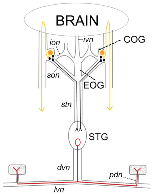
Figure 1.
The stomatogastric nervous system (STNS). Diagram of the STNS and connection to the brain. Nerves are drawn as lines and rectangles, muscles as stippled squares, dopamine-containing neurons as filled circles, and the pyloric dilator (PD) neuron as an open circle. The STNS comprises four ganglia: the two commissural ganglia (COGs), the esophageal ganglion (EOG), and the stomatogastric ganglion (STG). The PD neuron, located in the STG, projects an axon down the dorsal ventricular nerve (dvn). The axon bifurcates prior to entering the lateral ventricular nerves (lvn) and continues to run through the pyloric dilator nerves (pdn) to innervate the two PD muscles. Small DA-containing neurons in the COGs can project axons through the superior esophageal nerve (son) and down the stomatogastric nerve (stn) to terminate in the STG. The large L-cells in the COGs project to the ventral surface of the brain, whereupon axons reverse direction and ultimately terminate in ipsilateral pericardial organs, which release neuromodulators into the hemolymph (indicated by arrowheads). Not shown are the many dopaminergic cells in the brain (Tierney et al., 2003). In some species, neurons in the STG may also contain DA (Pulver et al., 2003). Dopamine has not been observed in the EOG, the inferior esophageal nerve (ion), or the inferior ventricular nerve (ivn) that connects the brain to the STNS.

