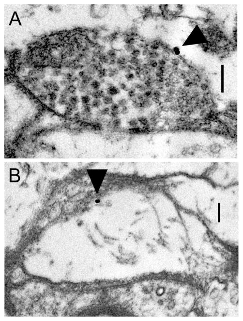
Figure 3.
D2αPan receptors can be located on presynaptic varicosities and terminal swellings. Electron micrographs showing anti-D2α immunogold staining (arrows) on a vesicle-containing varicosity (A) and a terminal swelling lacking vesicles (B). T-bars and postsynaptic densities indicative of synapses are not observed in these micrographs, suggesting that D2αPan receptors can be extrasynaptic. Scale bar = 0.25 μm in A,B.

