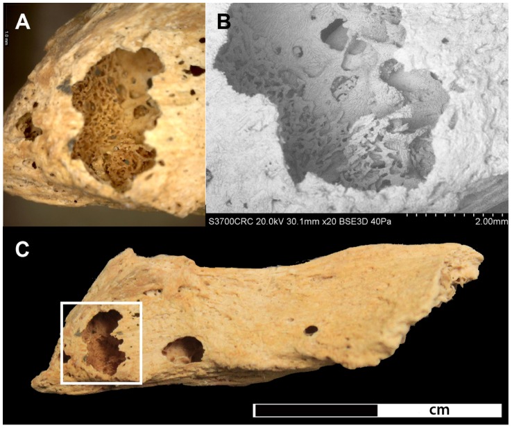
Figure 10. Lytic lesion in the spinous process of the 5th thoracic vertebra.
A) shows a close-up of bone formation at 35x magnification located within the lytic focus, B) SEM image of the lytic focus C) shows the complete spinous process with the location of the lytic focus highlighted in the rectangle.

