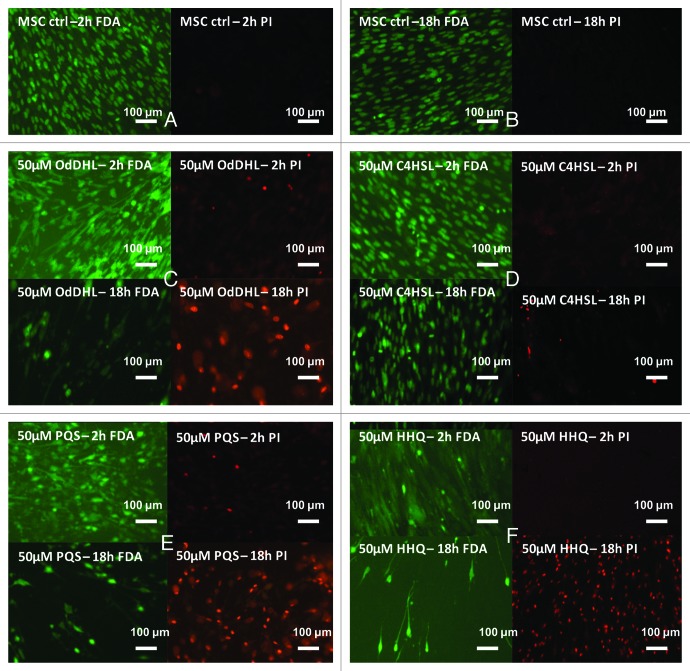
Figure 1. Fluorescence micrographs of MSCs monolayer grown in the presence of 50 μM purified QSSMs, for 2 and 18 h. (A and B) MSCs untreated control (containing an equivalent amount of HPLC grade EtOH), (C) MSCs treated with OdDHL, (D) MSCs treated with C4HSL, (E) MSCs treated with PQS, (F) MSCs treated with HHQ. FDA, fluorescein diacetate staining (green, visualization at 488 nm); PI, propidium iodide staining (red, visualization at 546 nm). Immersion oil, 1000× magnification.

An official website of the United States government
Here's how you know
Official websites use .gov
A
.gov website belongs to an official
government organization in the United States.
Secure .gov websites use HTTPS
A lock (
) or https:// means you've safely
connected to the .gov website. Share sensitive
information only on official, secure websites.
