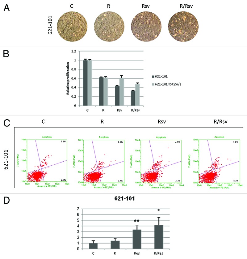
Figure 5. 621–101 cells show induction of apoptosis and decrease in proliferation upon combination rapamycin and resveratrol treatment. (A) 621–101 cells were treated with either 20 nM rapamycin and/or 100 µM resveratrol for 24 h. Cells were photographed using Zeiss light microscope under 10× magnification. (B) 621–101 and 621–101 TSC2 o/e cells were treated with 20 nM rapamycin and/or 100 µM resveratrol for 48 h, proliferation assay was performed as described in “Materials and Methods”. (C) 621–101 cells were treated as described in (A). Cells were subsequently scraped, pelleted and incubated with the Guava Nexin Reagent for 20 min at room temperature, and analyzed for Annexin V staining by flow cytometry. (D) 621–101 cells were treated as described in (A). Histogram represents quantification of early apoptotic cells from 3 experiments. Student’s t test was performed on treated samples relative to untreated controls. *P < 0.05, **P < 0.01.

An official website of the United States government
Here's how you know
Official websites use .gov
A
.gov website belongs to an official
government organization in the United States.
Secure .gov websites use HTTPS
A lock (
) or https:// means you've safely
connected to the .gov website. Share sensitive
information only on official, secure websites.
