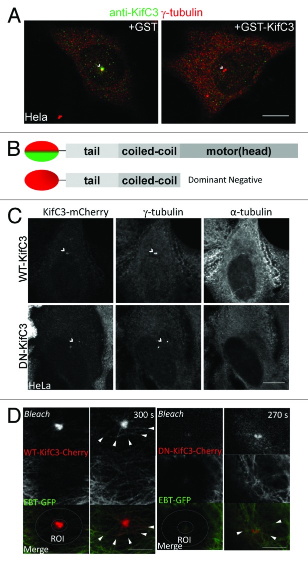
Figure 1. KIFC3 localizes to the centrosome in interphase. (A) HeLa cells were co-stained with anti-KIFC3 antibody (Lab antibody; green) and anti γ-tubulin (red). Primary antibody was incubated with either GST alone or GST-KIFC3 peptide antigen. Scale bar = 10 μm (B) Domain maps of WT-KIFC3 (top) and DN-KIFC3 with GFP or mCherry tag at the N terminus. (C) Microtubules were depolymerized in HeLa cells transfected with WT- or DN-KIFC3 mCherry (red) by incubation on ice and cells were stained with γ-tubulin (green) to visualize the centrosome and α-tubulin to confirm microtubule depolymerization (white). Scale bar = 10 μm (D) Photobleaching and recovery for MDCK cells expressing WT- or DN-KIFC3 (red) and EBT-GFP (green). Bleached area is indicated by a circle. Scale bar = 5 μm.
