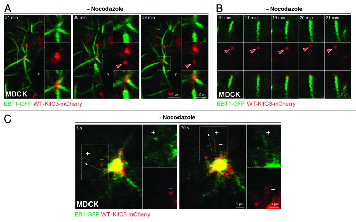
Figure 5. KIFC3 localizes to the minus-end of centrosomal-derived microtubules. (A and B) Time-lapse sequences that show growing microtubules in MDCK cells expressing WT-KIFC3-mCherry (red) and EBT1-GFP (green) after nocodazole washout. Red arrowheads indicate progressive movement of KIFC3-capped microtubules away from the centrosome (A) or in the cell periphery (B). n, nuclei. (C) Growing microtubules in MDCK cells expressing WT-KIFC3-mCherry (red) and microtubule plus-end protein EB1-GFP (green) after nocodazole washout. Note the segregation between KIFC3 and EB1 vesicular structures. “+” and “−” refer to the relative location of the presumed plus and minus-ends of a microtubule.

An official website of the United States government
Here's how you know
Official websites use .gov
A
.gov website belongs to an official
government organization in the United States.
Secure .gov websites use HTTPS
A lock (
) or https:// means you've safely
connected to the .gov website. Share sensitive
information only on official, secure websites.
