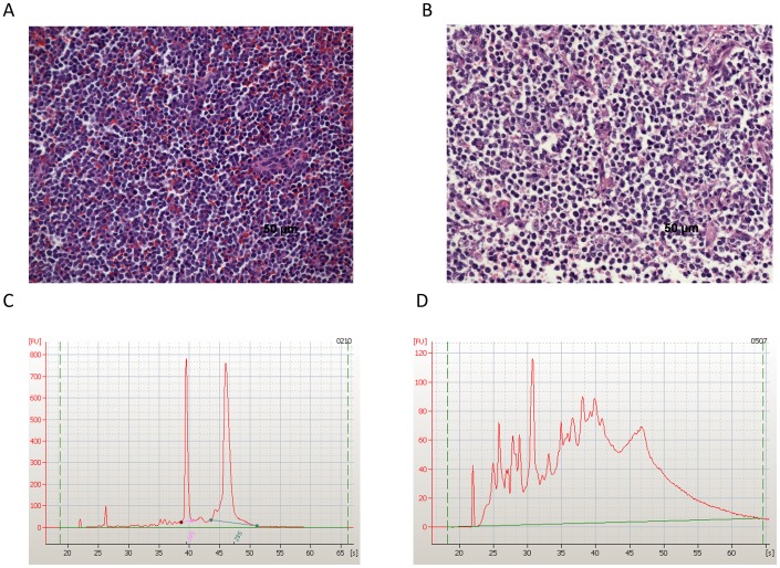
Figure 1. Histopathology and RNA quality assurance and control measures were successful in procuring high quality canine tumor samples.
Formalin-fixed, paraffin-embedded tumor biopsy samples were sectioned, paraffin embedded, and H&E stained for light microscopic evaluation. A single board-certified veterinary pathologist (EJE) assessed % tumor surface area, % tumor nuclei and % tumor necrosis to determine their quality prior to molecular profiling. Images of representative H&E images are shown: A. Sample 0209, a golden retriever with lymphoma, passed QA/QC. (Tumor 75–100%, necrosis <10%), while B. sample 0503, a beagle with lymphoma, failed QA/QC (Tumor 75–100%, necrosis >20%). Biopsies that failed to pass QA/QC in any category were excluded from subsequent analysis. Additionally RNA isolation was performed for all enrolled cases (n = 31) at a CLIA certified laboratory. RNA was extracted from Tumor A biopsy samples. Quality measures included quantity (total yield >20 ng) and integrity (A260/A280>1.8, RIN>8.0) measured by Nanodrop and Agilent Bioanalyzer. Electropherograms from cases C. 0210 and D. 0507 are depicted. Sample 0210, an oral melanoma, passed RNA QA/QC while sample 0507, a mast cell tumor, failed QA/QC (poor quality RNA due to a large connective tissue component).

