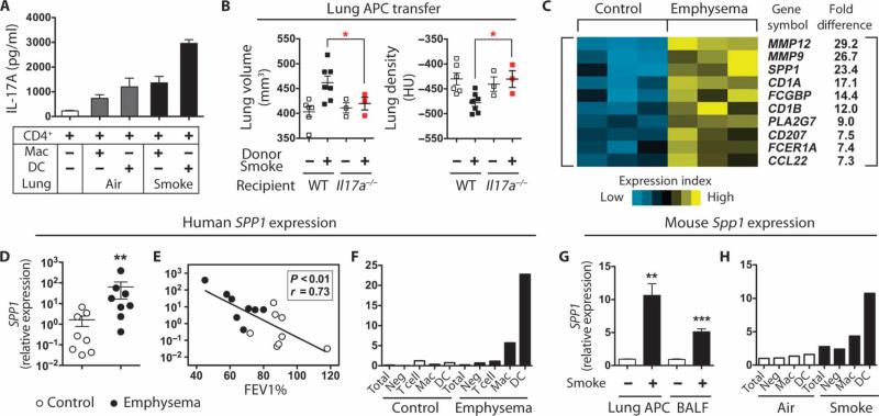
Fig. 3.
Mouse lung DCs induce TH17 differentiation. (A) Effect of CD4 T cells on IL-17A production in cells from air- or smoke-exposed mice. Splenic CD4 T cells were cultured with mouse lung macrophages (Mac) or DCs for 3 days with soluble anti-CD3 antibody (1 μg/ml), after which IL-17A was quantified from culture supernatants (n = 3). Data represent at least three independent studies. (B) μCT quantification of total lung volume and lung density from mice that received lung APCs from mice with the indicated phenotype (n = 3 to 7 per group). *P < 0.05. Data represent two independent studies. (C) Heat map depicting top 10 significantly up-regulated genes as determined by gene microarray analysis of human lung DCs from healthy control (n = 3) and emphysema (n = 3) subjects. (D) SPP1 mRNA expression in control lung DCs (n = 8) and emphysema lung DCs (n = 8). **P < 0.01. (E) Correlation of lung DC SPP1 mRNA expression and disease severity (FEV1%) (control, n = 8; emphysema, n = 8). (F) qPCR analysis of SPP1 mRNA expression in different lung cell population. Mac, macrophages; Neg, DC, macrophage, T cell–depleted cells. Data are representative of four independent experiments. (G) Spp1 mRNA expression in lung CD11c+ APCs and BALF cells from air-treated (n = 3) and cigarette smoking–treated (n = 3) mice. **P < 0.01; ***P < 0.001. (H) qPCR analysis of Spp1 mRNA expression in different lung cell population. Mac, macrophages; Neg, DC, macrophage-depleted cells. Data represent three independent studies with five mice pooled for each group.

