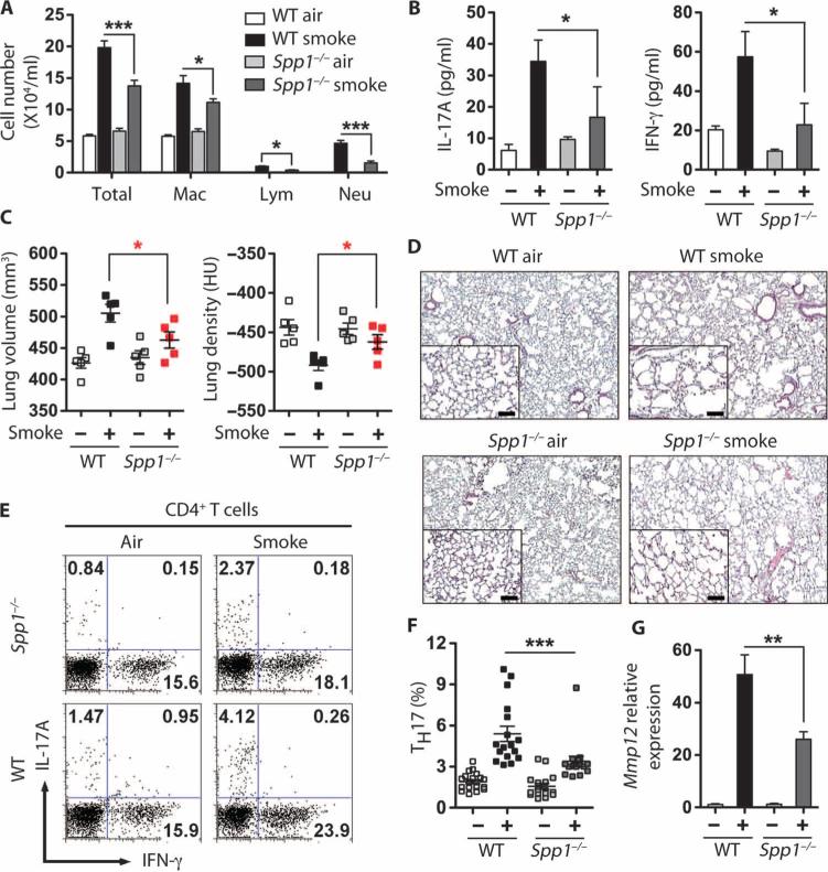
Fig. 5.
Spp1-deficient mice are protected from cigarette smoke–induced emphysema. (A) Number of cells in BALF of air- and cigarette smoke–exposed mice. Total cells, macrophages (Mac), lymphocytes (Lym), and neutrophils (Neu) (n = 5 per group). *P < 0.05; ***P < 0.001. (B) IL-17A and IFN-γ levels in cells from airand cigarette smoke–exposed Spp1−/− mice. CD11c-depleted cells from whole lung homogenates were stimulated overnight with PMA and ionomycin, and supernatant protein concentrations were measured. n = 3 to 5 per group. *P < 0.05. (C) μCT quantification of total lung volume and lung density from air (–)– and cigarette smoke (+)–exposed WT and Spp1−/− mice (n = 5 per group). *P < 0.05. Data represent at least three independent studies. (D) Representative H&E staining of lung sections from WT and Spp1−/− mice exposed to air or smoke for 4 months; insets represent ×200 magnification. Scale bars, 100 μm. (E and F) Representative (E) and cumulative (F) intracellular cytokine analyses of CD4 T cells derived from lungs of air- and smoke-exposed WT and Spp1−/− mice (n = 16 to 20). ***P < 0.001 by one-way ANOVA test. (G) Mmp12 mRNA expression in total BALF cells from air- and smoke-exposed mice (n = 5 per group). **P < 0.01.

