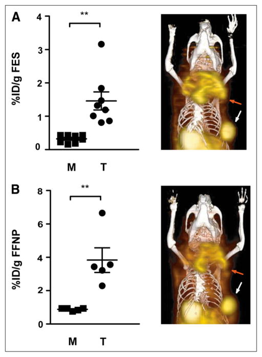
FIGURE 1.
Aged female STAT1−/− mice with primary mammary tumors were imaged with small-animal PET/CT using 18F-FES (7 mice; 8 tumors) (A) and 18F-FFNP (4 mice; 5 tumors) (B). Activity in tumor and muscle was measured and graphed. Coronal 3-dimensional fused small-animal PET/CT images are shown for mouse with large primary tumor in left upper thoracic fat pad (red arrow) and smaller tumor in left lower thoracic fat pad (white arrow). Intense physiologic activity is present in gallbladder and bowel, consistent with hepatobiliary clearance. **P < 0.01.

