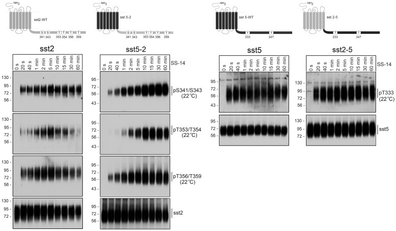
Figure 1. Agonist-induced phosphorylation of sst2 and sst5 tail-swap mutants.
Top, Schematic representation of the human wild-type sst2 (depicted in grey) and human wild-type sst5 receptors (depicted in black) and their corresponding tail-swap mutants. Phosphate acceptor sites targeted for the generation of phosphosite-specific antibodies are depicted as circles. Bottom, stably transfected HEK 293 cells were exposed to 1 µM SS-14 at room temperature for the indicated time periods. Cells were lysed and immunoblotted with the indicated phosphosite-specific antibodies. Blots were then stripped and reprobed with the phosphorylation-independent anti-sst5 antibody {UMB-4} or anti-sst2 antibody {UMB-1} to confirm equal loading of the gels. Shown are representative results from one of three independent experiments. The position of molecular mass markers is indicated on the left (in kDa).

