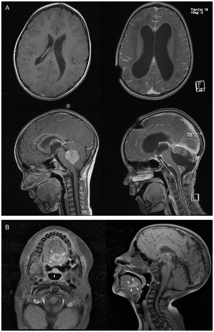
Figure 3. MR imaging documenting severe normal tissue toxicity or synchronous tumors in patients.
(A) Cranial magnetic resonance imaging of the patient GRJN before (left panel) and after tumour resection and adjuvant radio-/chemotherapy (right panel). T1-weighted images acquired in sagittal and axial plane shows cerebral atrophy with enlargement of the ventricles after multifactorial leukencephalopathy. (B) Cranial magnetic resonance imaging of the patient SBNE before surgical tumor resection. T1-weighted images acquired in sagittal and axial plane reveal the squamous cell carcinoma (SCC) of the oral cavity (tumors marked by arrows).

