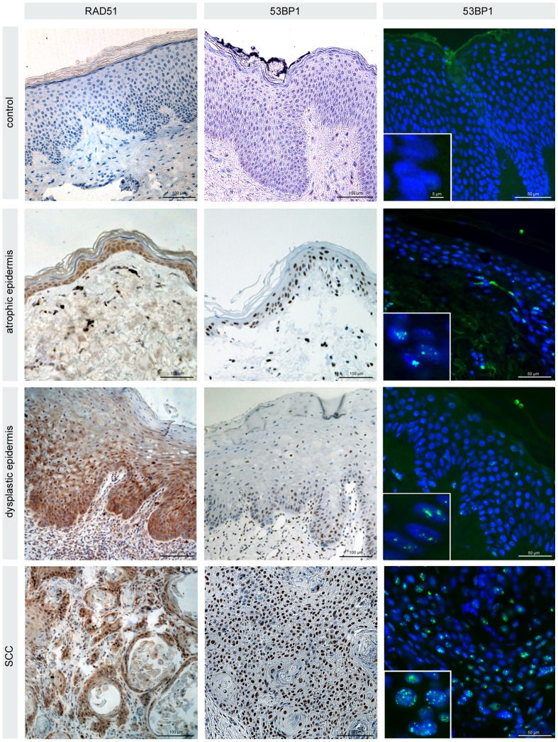
Figure 4. Light microscopy analysis of the skin and tumor obtained from the Fanconi patient.
Immunohistochemical staining of RAD51 and 53BP1 (diaminobenzidine, brown) in atrophic and dysplastic epidermis as well as tumor specimens derived from the Fanconi patient. Compared with healthy control epidermis moderately or considerably increased RAD51 and 53BP1 expression in the proliferating zone attached to the basement membrane. Immunfluorescence staining of 53BP1 (green) reveals discrete nuclear foci in the proliferating cells of the epidermis, with increasing foci levels during tumor development. Original magnification, x600.

